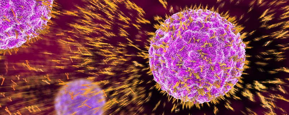
My Wishlist

View your shopping bag

Items: (0) £0.00
  • £
  • $
  • €

Checkout

Cancer

  • Grid View
  • List View
3D1.1 Antibody (Parathyroid hormone like protein) - Mouse Anti PTHLH
The protein encoded by this gene is a member of the parathyroid hormone family. This hormone regulates endochondral bone development and epithelial-mesenchymal interactions during the formation of the mammary glands and teeth. This hormone is involved in lactation possibly by regulating the mobilization and transfer of calcium to the milk. The receptor of this hormone, PTHR1, is responsible for most cases of humoral hypercalcemia of malignancy. Four alternatively spliced transcript variants encoding two distinct isoforms have been observed. There is also evidence for alternative translation initiation from non-AUG (CUG and GUG) start sites, in-frame and downstream of the initiator AUG codon, to give rise to nuclear forms of this hormone
£226.00

Buy | View ›

Adrenocorticotropic hormone (ACTH) N-Terminal Antibody - Mouse Anti-Adrenocorticotropic hormone (ACTH) N-Terminal
ACTH occurs in cells of the anterior pituitary and in neurons in brain. It regulates the corticosteroid production in the adrenal cortex. Beta endorphin and Met enkephalin are endogenous opiates. MSH (melanocyte stimulating hormone) increases the pigmentation of skin by increasing melanin production in melanocytes
£226.00

Buy | View ›

Apolipoprotein E4 Antibody ; Mouse Anti-Apolipoprotein E4
Apolipoprotein E, a main apoprotein of the chylomicron, binds to a specific receptor on liver cells and peripheral cells and is essential for the normal catabolism of triglyceride-rich lipoprotein constituents.ApoE exists in three major isoforms; E2, E3, and E4, which differ from one another by a single amino-acid substitution. Individuals heterozygous for the ApoE4 allele are at higher risk of late-onset Alzheimer's disease. Defects in apolipoprotein E result in familial dysbetalipoproteinemia, or type III hyperlipoproteinemia (HLP III), in which increased levels of plasma cholesterol and triglycerides are the consequence of impaired clearance of chylomicron and VLDL remnants
£226.00

Buy | View ›

Aurora B Antibody - Sheep Anti- Aurora B 0.2ml
Aurora B is a mitotic protein kinase that phosphorylates histone H3 (probably on Serine 10) behaves as a chromosomal passenger protein and may regulate several stages of mitosis such as centrosome separation chromosome segregation and cytokinesis It localizes to the inner centromere region from prophase to anaphase
£226.00

Buy | View ›

BUB1 Antibody - Mouse (monoclonal) Anti-BUB1 0.1ml
Bub1 is a kinase involved in spindle checkpoint function The kinase functions in part by phosphorylating a member of the miotic checkpoint complex and activating the spindle checkpoint Mutations in Bub1 have been associated with aneuploidy and several forms of cancer
£226.00

Buy | View ›

BUB1 Antibody - Mouse Anti-BUB1 50ul
Bub1 is a kinase involved in spindle checkpoint function The kinase functions in part by phosphorylating a member of the miotic checkpoint complex and activating the spindle checkpoint Mutations in Bub1 have been associated with aneuploidy and several forms of cancer
£183.00

Buy | View ›

Cytokeratin (keratin) K13 Antibody (AE8) - Mouse Anti-Cytokeratin (keratin) K13, AE8 0.1ml
Cytokeratin 13 is a member of the keratin gene family The keratins are intermediate filament proteins responsible for the structural integrity of epithelial cells and are subdivided into cytokeratins and hair keratins Most of the type I cytokeratins consist of acidic proteins which are arranged in pairs of heterotypic keratin chains This type I cytokeratin is paired with keratin 4 and expressed in the suprabasal layers of non-cornified stratified epithelia Mutations in this gene and keratin 4 have been associated with the autosomal dominant disorder White Sponge Nevus This antibody is specific for Cytokeratin 13 which is a marker for oesophageal type differentiation which is expressed by various internal stratified epithelia
£226.00

Buy | View ›

EAPP (E2F- associated phosphoprotein) Antibody ; Mouse Anti-EAPP (E2F- associated phosphoprotein)
Transcription activator that binds DNA cooperatively with dp proteins through the E2 recognition site, 5'-TTTC[CG]CGC-3' found in the promoter region of a number of genes whose products are involved in cell cycle regulation or in DNA replication. The DRTF1/E2F complex functions in the control of cell-cycle progression from G1 to S phase. E2F-1 binds preferentially RB1 protein, in a cell-cycle dependent manner. It can mediate both cell proliferation and p53-dependent apoptosis
£226.00

Buy | View ›

EPCAM; CD326 Antibody- Rat Anti-EPCAM
Epithelial Cell Adhesion Molecule (EpCAM) is a 40 kDa cell surface antigen. This antigen has been identified independently by a number of groups, and has been known by a variety of names. Several monoclonal antibodies have been raised against EpCAM, many of which have been described as tumour specific molecules on carcinomas. EpCAM is a Type 1 transmembrane glycoprotein. It is expressed on the basolateral membrane of cells by the majority of epithelial tissues, with the exception of adult squamous epithelium and some specific epithelial cell types including hepatocytes and gastric epithelial cells. EpCAM expression has been reported to be a possible marker of early malignancy, with expression being increased in tumour cells, and de novo expression being seen in dysplastic squamous epithelium
£226.00

Buy | View ›

FNIP1 folliculin interacting protein 1 Antibody- Rabbit Anti-FNIP1
Birt-Hogg-Dubé syndrome, a hamartoma disorder characterized by benign tumors of the hair follicle, lung cysts, and renal neoplasia, is caused by germ-line mutations in the BHD(FLCN) gene, which encodes a tumor-suppressor protein, folliculin (FLCN), with unknown function. The tumor-suppressor proteins encoded by genes responsible for several other hamartoma syndromes, LKB1, TSC1/2, and PTEN, have been shown to be involved in the mammalian target of rapamycin (mTOR) signaling pathway
£226.00

Buy | View ›

HCG beta Antibody- Mouse anti-HCG beta
Human chorionic gonadotropin (hCG) is a glycoprotein hormone produced by trophoblastic cells of the placenta beginning 10 to 12 days after conception. Maintenance of the fetus in the first trimester of pregnancy requires the production of hCG, which binds to the corpus luteum of the ovary which is stimulated to produce progesterone which in turn maintains the secretory endometrium. The unique beta chain confers biological specificity to thyrotropin, lutropin, follitropin and gonadotropin. hCG acts to maintain the corpus luteum until the developing placenta is able to produce the required levels of oestrogen and progesterone.
£226.00

Buy | View ›

HDAC3 Antibody ; Mouse Anti-HDAC3
Responsible for the deacetylation of lysine residues on the N-terminal part of the core histones (H2A, H2B, H3 and H4). Histone deacetylation gives a tag for epigenetic repression and plays an important role in transcriptional regulation, cell cycle progression and developmental events. Histone deacetylases act via the formation of large multiprotein complexes. Probably participates in the regulation of transcription through its binding to the zinc-finger transcription factor YY1; increases YY1 repression activity. Required to repress transcription of the POU1F1 transcription factor. Acts as a molecular chaperone for shuttling phosphorylated NR2C1 to PML bodies for sumoylation
£226.00

Buy | View ›

HEF 1/Cas-L (NEDD9)Antibody - Mouse Anti-HEF-1/Cas-L
HEF1 is a multifunctional protein involved in integrin-based signaling that affects cell motility growth apoptosis and oncogenic transformation The Cas family of docking proteins have been the subject of intense research because of their role in cell motility growth apoptosis and oncogenic transformation These proteins are substrates of focal adhesion kinase (FAK) and the Src family of tyrosine kinases two active targets for drug development HEF1 protein production increases levels of mRNA transcripts that encode proteins associated with motility cell transformation and invasiveness including several metalloproteinases MLCK p160ROCK and ErbBi HEF1 overproduction also mediates apoptosis in epithelial-derived cell lines including MCF7 and HeLa cells Recent clinical studies at another institution have found that overexpression of BCAR1 (p130Cas) a related protein is associated with tamoxifen resistance This highlights the importance of studying the role of this family of proteins in cancer prognosis
£226.00

Buy | View ›

HEF-1/ Cas-L Antibody- Mouse Anti-HEF-1/ Cas-L
HEF1 is a multifunctional protein involved in integrin-based signaling that affects cell motility growth apoptosis and oncogenic transformation The Cas family of docking proteins have been the subject of intense research because of their role in cell motility growth apoptosis and oncogenic transformation These proteins are substrates of focal adhesion kinase (FAK) and the Src family of tyrosine kinases two active targets for drug development HEF1 protein production increases levels of mRNA transcripts that encode proteins associated with motility cell transformation and invasiveness including several metalloproteinases MLCK p160ROCK and ErbBi HEF1 overproduction also mediates apoptosis in epithelial-derived cell lines including MCF7 and HeLa cells Recent clinical studies at another institution have found that overexpression of BCAR1 (p130Cas) a related protein is associated with tamoxifen resistance This highlights the importance of studying the role of this family of proteins in cancer prognosis
£226.00

Buy | View ›

Lamin A/C mutant R453W Antibody ; Mouse Anti-Lamin A/C mutant R453W
Nuclear lamins are intermediate filament proteins that are the major structural component of the nuclear lamina on the inner surface of the nuclear envelope. Lamin A and Lamin C are splice variants of the Lamin A gene. Lamin A/C (CDCD1, LMN1, EMD2) expression is a hallmark of embryonic stem cell differentiation. In addition to adding structural integrity to the nucleus, lamins contribute to the makeup of the nuclear matrix. Lamins also help organize interphase chromatin through interactions with several chromatin proteins, including histones and Lap2, such that alteration in lamin organization (laminopathy) results in disruption of DNA replication, transcription and RNA processing. The R453W mutation is one of the most common causes of autosomal dominant Emery-Dreifuss muscular dystrophy (EDMD)
£226.00

Buy | View ›

Lamin C Antibody- Rabbit Anti-Lamin C
An important part of the cell nucleus is formed by nuclear lamina Nuclear lamins form a network of filaments at the nucleoplasmic site of the nuclear membrane Two main subtypes of nuclear lamins can be distinguished ie A-type lamins and B-type lamins The A-type lamins comprise a set of three proteins arising from the same gene by alternative splicing ie lamin A lamin C and lamin Adel10 while the B-type lamins include two proteins arising from two distinct genes ie lamin B1 and lamin B2 The nuclear lamins comprise a unique subclass of the intermediate filament protein family They share a molecular domain organisation with the other intermediate filament proteins in that they are fibrous molecules that have an aminoterminal globular head a central rod of a-helices and a carboxyterminal globular domain Many biochemical and molecular features of lamins have been studied but their functions remain still largely undetermined One of the functions ascribed to the lamina is the maintenance of the structural integrity of the nucleus Besides interactions with the nuclear membrane and other intermediate filaments lamins interact with the nuclear chromatin Eukaryotic chromatin is organised into loops which are attached to the nuclear matrix This organisation is thought to contribute to compaction of the chromatin and regulation of gene expression Lamins as part of the nuclear matrix may be involved in these processes since chromatin binding sites have been detected in both A- and B-type lamins
£226.00

Buy | View ›

View: 16 / 32 / 48 / 64 / 80 / products per page

« previous 1 2 3 4 5 6 next »