Cell Cycle Antibodies for research into the Cell Cycle which has four distinct phases, G1, S, G2 and M collectively representing cell division which follows a highly regulated pathway culminating in mitosis in eukaryotes.
Cell cycle antibody research into disruption of the cell cycle has been implicated in tumour formation and growth, active cell division targeting in treating cancer and negative cardiovascular incidents.
Cell cycle antibodies are available in the following volumes including 0.05 ml, 0.1ml, 0.2 ml, 1 ml and 2 ml.
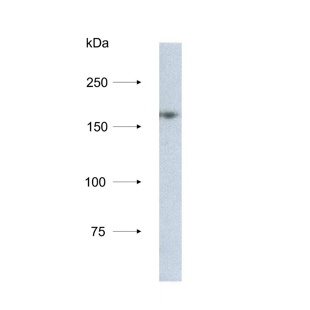
NCAPD3/hCAP-D3 Monoclonal Antibody [2B5]
| Product Code: | IQ554 |
| Reactivity: | Human |
| Application: | WB, IP |
| Citation Count: | 2 |
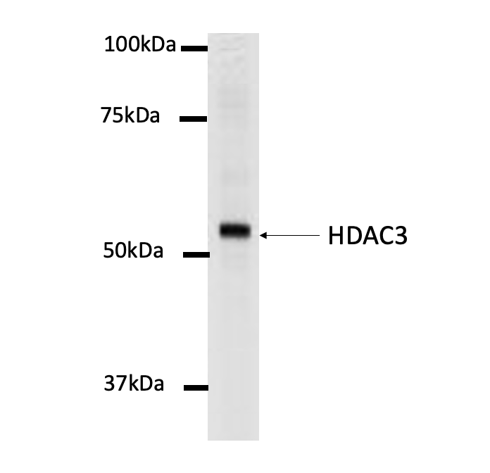
HDAC3 Monoclonal Antibody [3G6]
| Product Code: | IQ553 |
| Reactivity: | Human, Mouse |
| Application: | WB , IP, ICC/IF |
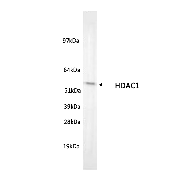
HDAC1 Monoclonal Antibody [10E2]
| Product Code: | IQ555 |
| Reactivity: | Mouse, Rat, Human |
| Application: | Flow Cyt, WB, ICC/IF, IP, ChIP, IHC-P |
| Citation Count: | 1 |